Follicular Unit Extraction Technique Explained: The Anatomy-First Breakdown
Introduction: Why Most FUE Explanations Fall Short
Most patient-facing content about follicular unit extraction describes what the procedure is without explaining why each technical decision is made. This gap leaves prospective patients without a genuine framework for evaluating surgeon skill and clinic quality.
Follicular unit extraction—or more accurately, follicular unit excision (FUE)—is a minimally invasive hair transplant technique in which individual follicular units, the natural groupings of one to four hairs, are extracted one at a time from the donor area and implanted into thinning or balding recipient zones. The International Society of Hair Restoration Surgery (ISHRS) formally updated the terminology from “extraction” to “excision” to better reflect the operative decision-making and surgical skill the procedure genuinely requires. This distinction, absent from nearly all competitor content, signals something important: FUE is not a simple harvesting exercise but a precise surgical intervention governed by anatomy at every step.
This article anchors every procedural step to the underlying anatomy that governs it—from punch size selection to graft preservation chemistry. At Hair Doctor NYC, the philosophy centers on treating patients as educated partners in their own care, and that begins with transparency about the clinical reasoning behind every decision.
The Anatomy That Drives Every FUE Decision
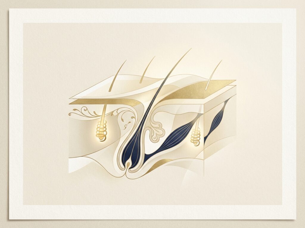
A follicular unit is a naturally occurring grouping of one to four terminal hairs, a sebaceous gland, an arrector pili muscle, and associated connective tissue—all enclosed in a shared adventitial sheath. Understanding this structure is essential because every downstream technical decision responds directly to these anatomical realities.
The follicle contains three anatomical zones with different mechanical properties relevant to extraction. The infundibulum comprises the upper segment extending from the skin surface to the sebaceous gland opening. The isthmus represents the middle segment where the arrector pili muscle attaches—this is the anatomically tightest point of the follicle’s connection to surrounding tissue and the zone most vulnerable to transection during punch penetration. The inferior segment contains the bulb region where active hair growth occurs.
Follicular angulation presents the first technical challenge. Scalp follicles grow at an acute angle beneath the skin surface, not perpendicular to it. The punch must be aligned with the follicle’s subsurface trajectory, not just its visible surface exit angle. Compounding this challenge is follicular curvature: follicles are not straight tubes but slightly curved structures. This curvature increases transection risk if the punch is advanced too deeply or at the wrong angle.
Patients with Afro-textured, curly, or coily hair face significantly higher transection risk because their follicle paths curve more dramatically beneath the skin. These patients may require specialized techniques, larger punches, or consideration of FUT as an alternative approach.
The FOX Test: Anatomy-Based Pre-Operative Suitability Assessment
The FOX test (Follicular Unit Extraction test) is a pre-operative suitability assessment in which the surgeon extracts approximately 100 grafts from the donor area and evaluates the transection rate before committing to a full procedure.
The anatomical rationale is straightforward: because follicle angulation, curvature, and skin laxity vary significantly between patients, a test extraction reveals whether a given patient’s follicular architecture is amenable to FUE without unacceptable graft loss. A passing result demonstrates that the surgeon can extract grafts cleanly; a failing result may indicate the patient would be better served by FUT.
The FOX test also helps the surgeon calibrate the optimal punch size and technique for that specific patient’s anatomy before the full session begins. Its near-total absence from competitor patient-education content represents a significant transparency gap—and its presence in a clinic’s protocol signals rigorous, individualized anatomical evaluation.
Punch Size Selection: The Anatomy-First Calculation
FUE punches range from 0.6mm to 1.0mm in diameter, and punch size selection is a direct calculation based on the follicular unit’s cross-sectional diameter at the level of the arrector muscle attachment.
The trade-off is precise: a punch that is too small risks transecting the follicle; a punch that is too large removes excess surrounding tissue, creates larger wounds, increases scarring risk, and reduces donor area density. Hair caliber (fine versus coarse), follicular grouping size (singles versus quadruples), and skin type all influence the optimal punch diameter.
Smaller punches (0.6–0.75mm) are associated with less visible scarring and faster healing but demand greater technical precision. These are typically used for fine-haired patients or in high-density donor areas.
Larger punches (0.9–1.0mm) may be necessary for coarser hair or multi-hair follicular units but require careful donor area management to avoid a “moth-eaten” appearance from overharvesting.
In high-volume sessions exceeding 3,000–4,000 grafts, even a slightly oversized punch multiplied across thousands of extraction sites can compromise donor area integrity. The ARTAS robotic system addresses this by using 44-micron precision imaging to optimize punch placement, reducing the human error margin in sizing and positioning.
Sharp vs. Blunt Punch Mechanics: Two-Step and Three-Step Techniques Explained
The choice between sharp and blunt punches is not a matter of preference but of anatomical strategy.
The two-step technique uses a sharp punch to score through the epidermis and dermis in a single motion, severing the follicle’s connective tissue attachments before the graft is removed with forceps. The advantage is speed; the risk is a higher transection rate if depth control is imprecise.
The three-step technique, exemplified by the SAFE System (Surgically Advanced Follicular Extraction), proceeds differently. Step one uses a sharp punch to score only the epidermis. Step two uses a blunt punch to dissect through the dermis and around the follicle without cutting it—the blunt punch’s rounded tip pushes tissue aside rather than cutting it, dramatically reducing transection risk at the critical arrector muscle attachment zone. Step three removes the graft.
The three-step technique is anatomically superior for reducing transection because the blunt punch cannot accidentally sever the follicle at the tight isthmus zone. The trade-off is that it requires more time and greater surgical skill.
Motorized and oscillating devices interact with these mechanics in important ways. Oscillating punches reduce the torque applied to the follicle during penetration, which is particularly beneficial for curved follicles. Under 2.5–5× magnification, surgeons can visually confirm follicular alignment before committing the punch to full depth.
The Extraction Tool Spectrum: From Manual Punch to AI-Guided Robotics
- Manual punch: The original FUE instrument (0.7–1.0mm hollow scalpel) requiring the surgeon to manually control depth, angle, and rotation. Highest skill dependency with no automation buffer against human error.
- NeoGraft: Motorized rotating punch with suction-assisted graft removal. Reduces manual fatigue in large sessions, though the suction mechanism can stress grafts if vacuum pressure is improperly calibrated.
- SmartGraft: Suction-based system with touchscreen-controlled temperature storage for extracted grafts, directly addressing the graft preservation challenge during long sessions.
- ARTAS Robotic System: FDA-cleared system using 3D optical imaging and AI to identify follicular units, calculate optimal punch angle and depth, and execute extractions with 44-micron precision at 500–700 grafts per hour.
- HARRTS FUEsion X 5.0: A system integrating AI, robotics, and augmented reality for real-time scalp mapping and graft distribution planning, representing a current frontier of extraction technology.
No tool eliminates the need for surgical judgment. The ISHRS has issued warnings about unlicensed technicians performing extractions—a patient safety concern underscoring why the surgeon’s role in tool selection and oversight remains non-negotiable.
Recipient Site Creation: The Artistic and Anatomical Precision Step
Recipient site creation is frequently glossed over in patient-facing content despite being one of the most technically demanding and artistically consequential steps in the procedure.
The surgeon creates tiny incisions in the recipient area where grafts will be placed, calculating four critical variables for each: depth (must match the graft length being implanted), diameter (must match the graft’s cross-sectional width), angle (typically 45° to mimic natural hair growth direction), and direction (must align with the surrounding hair’s natural growth pattern).
Sapphire FUE uses sapphire-tipped blades that allow finer, more precise incisions than steel blades, reducing tissue trauma, minimizing edema, and enabling higher-density implantation. DHI (Direct Hair Implantation) with the Choi Implanter Pen combines site creation and graft implantation in a single step, reducing graft out-of-body time.
The surgeon’s aesthetic judgment—hairline design, density distribution, directional variation—is exercised throughout this step. A background in facial plastic surgery provides meaningful advantage here, as understanding facial harmony and aesthetic proportion is as important as technical precision.
Graft Preservation: The Out-of-Body Science That Determines Survival
Once a follicular unit is extracted, it begins experiencing ischemic stress. Four primary threats endanger extracted grafts: dehydration of the outer root sheath, temperature elevation causing metabolic acceleration and cellular damage, mechanical trauma from compression during handling, and ischemia from oxygen and nutrient deprivation.
Cold hypothermic storage in chilled saline or specialized nutrient solutions slows cellular metabolism, extending viable out-of-body time. This is especially critical in mega-sessions where some grafts may remain out of body for several hours. Advanced preservation solutions such as HypoThermosol provide ionic balance and antioxidant protection that plain saline cannot offer.
Some clinics add PRP (platelet-rich plasma) to the storage solution, bathing grafts in growth factors during out-of-body time to potentially improve post-implantation survival. Reputable clinics using advanced techniques report 90–95% graft survival rates; scalp hair averages 89% survival, beard hair 95%, and body and chest hair approximately 76% at one year.
The Complete FUE Procedure Sequence: Step-by-Step With Anatomical Rationale
Step 1: Consultation, Donor Assessment, and Safe Donor Area Mapping
The safe donor area (SDA) encompasses the occipital and parietal regions where follicles are genetically resistant to DHT-driven miniaturization. Trichoscopy-guided donor assessment evaluates follicular density, miniaturization ratio, and hair caliber before planning extraction volume. Overharvesting beyond the SDA’s sustainable yield creates a “moth-eaten” appearance that is difficult to correct.
Step 2: Donor Area Preparation and Anesthesia
The donor area is trimmed to 1–2mm to allow visualization of follicular exit angles. Tumescent anesthesia creates a firm tissue plane that stabilizes follicles during extraction, reduces bleeding, and slightly separates the follicle from surrounding tissue.
Step 3: Follicular Unit Extraction
The punch is aligned with the follicle’s surface exit angle, advanced to the appropriate depth, and rotated or oscillated to sever connective tissue attachments. Depth control is critical: too shallow leaves the graft attached; too deep risks transecting the bulb. Grafts are harvested in a dispersed pattern across the donor area to maintain density.
Step 4: Graft Sorting, Inspection, and Preservation
Extracted grafts are inspected under magnification and sorted by follicular unit size for strategic placement—single-hair grafts at the hairline for natural softness, multi-hair grafts in the mid-scalp and crown for density.
Step 5: Recipient Site Creation and Graft Implantation
Recipient sites are created with attention to angle, direction, depth, and density calibrated to scalp vascularity. Grafts are implanted using fine forceps or a Choi implanter pen, with the surgeon’s aesthetic judgment shaping the final result.
Recovery Timeline and the Biology of Hair Regrowth
Understanding the regrowth timeline requires understanding the hair growth cycle. Days 1–7 bring swelling, mild discomfort, and crusting; most patients return to normal activities within 3–7 days. Weeks 2–4 see shock loss as transplanted hairs shed—a normal, anatomically expected response. Months 3–4 bring initial regrowth as follicles re-enter the anagen phase. Months 6–9 show progressive thickening, with full results visible at months 9–18.
FUE leaves only tiny dot-like white scars (0.6–1.0mm) rather than FUT’s linear scar, making it ideal for patients who prefer shorter hairstyles.
FUE vs. FUT: When Anatomy Determines the Better Choice
FUE and FUT are different anatomical approaches to the same goal. FUE advantages include no linear scar, faster recovery, the ability to harvest from non-scalp donor areas, and suitability for patients wearing short hair. FUT advantages include higher graft yield per session, lower transection rate (follicles are dissected under direct visualization), and preservation of donor surface integrity for future FUE sessions.
FUE holds 58–70% global market share, reflecting patient preference for minimal scarring rather than universal technical superiority for every case.
What Separates a Technically Excellent FUE Clinic From an Average One
Key quality indicators include: pre-operative FOX testing demonstrating individualized assessment; punch selection transparency in which the surgeon explains the rationale; proper magnification and graft handling protocols; surgeon-performed extractions rather than unsupervised technicians; recipient site artistry informed by facial aesthetics training; and documented graft preservation protocols.
Hair Doctor NYC exemplifies these standards with Dr. Roy B. Stoller (double board-certified, 25+ years of experience, 6,000+ procedures), Dr. Louis Mariotti (double board-certified facial plastic surgeon), and Dr. Christopher Pawlinga (18 years exclusively dedicated to hair transplantation).
Conclusion: Anatomy Is the Foundation — Surgeon Skill Is the Structure
Every technical decision in FUE—from punch diameter to preservation chemistry to recipient site angle—responds directly to follicular anatomy. The ISHRS terminology shift from “extraction” to “excision” reflects the surgical judgment, anatomical knowledge, and technical precision the procedure requires.
FUE’s dominance as the most widely performed hair transplant technique globally reflects genuine clinical advantages—but those advantages are realized only when the procedure is performed with the anatomical rigor described here. The FOX test, punch selection rationale, graft preservation protocols, and recipient site planning are the questions patients should raise at any consultation.
Ready to Understand Your Options With a Surgeon Who Will Explain Every Step?
Hair Doctor NYC invites prospective patients to schedule a consultation where the surgical team will apply this anatomical framework to each patient’s specific hair loss pattern, donor area characteristics, and aesthetic goals. The consultation is an educational conversation—patients receive a candid assessment of their FUE candidacy, including whether a FOX test is indicated and what graft yield is realistically achievable.
Multiple board-certified surgeons with decades of specialized experience practice at the state-of-the-art clinic on Madison Avenue in Midtown Manhattan. Contact Hair Doctor NYC to schedule a personalized consultation and take the first step toward a technically informed hair restoration decision.
Excellence Meets Elegance—surgical precision combined with a premium, personalized patient experience.